(Click here to read our disclaimer)
(Click here to buy Mgf in our store)
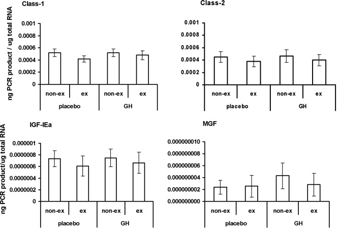
Mgf refers to mechano growth factor, a hormone that is produced in muscle tissue of animals when the muscles are trained. This chemical is considered to be an isoform of IGF-1. This spliced variant shares the mature region with IGF-1 but has a different E domain. The C-terminal sequence between these two chemicals shows a distinct variant that allows researchers to differentiate between the two bodies.
Mgf is associated with promoting healing in the muscles of animal subjects after they have been damaged. In a natural setting, the chemical can stimulate the stem and satellite cells in addition to the necessary anabolic processes to promote the growth of new tissue or muscle fibers by tissues that were not damaged.
It is believed that mgf and IGF-1 are central to the regrowth of muscle and the primary anabolic mechanism used to create new tissue.
Inability to Naturally Express Mgf Due to Mechanical Overload
Studies are investigating the response of IGF-1 signals and the capacity of skeletal muscle in an animal subject to react to the mechanical overload of this chemical by using synergistic muscle ablation.
- Overloading the soleus and plantaris caused a significant hypertrophy response as well as activation of the satellite cells. This response was more noticeable in younger rats.
- To splice variants of IGF-1 were found to be regulated differentially by RNA levels.
Significant changes that could be associated with an inability of older muscles in the subject’s body to manage mechanical overload resulted in a lower expression of the splice variant applied in that area as well as the machano growth factor.
This also created a failure for these muscles to un-regulate the IGF-1 receptors, mRNA and MyoD.
Expression of Actions in Rat Cardiomyocytes
A study worked to investigate the expression patterns of IGF-1 gene in rat myocardium between one hour and eight weeks after an induced myocardial infarction that was caused by coronary artery ligation at left anterior.
- Mgf-E and IGF-1 actions were characterized as well as the respective signals to H9C2 myocardial-like cells that were in vitro.
- Throughout the study period mgf and IGF-1Ea expressions increased significantly at the translational and transcriptional levels, particularly during the post-infarction period at 4-8 weeks.
- The measurements of IGF-1 serum levels in the rats decreased in the 24 hour to one week period, but then did not change in the later portions of the experiment when they were compared with sham-operated control rats.
- The specific anti-IGF-1r neutralizing antibodies during this period failed to block synthetic mgf-E particles and did not activate the Akt phosphorylation, but groups exposed to ERK1/2 did.
The summary of this data leads researchers to believe that the expression of IGF-1 during myocardial repair is essential. It also leads researchers to believe that the presence of synthetic mgf-E peptides could be mediated using an IGF-1R independent pathway, as was expressed in the in-vitro experiments focusing on the rat myocardial cells.
Continuing research is necessary to determine how mgf varies from other naturally occurring peptides as well as how to naturally stimulate this chemical in animals.
Sources:
http://www.sciencedirect.com/science/article/pii/S0014579301028253
http://www.ncbi.nlm.nih.gov/pmc/articles/PMC2656994/
Click here to view our entire PDF research library
Click here to view/download the PDF version of this article