What is Melanotan II?

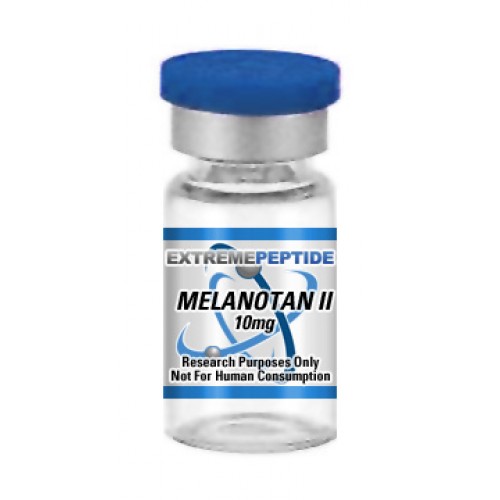
Melanotan II, also known as Melanotan 2 or MT-2 but different from Melanotan I, is a peptide that has a molecular mass of 1024.180 and a molecular formula of C50H69N15O9.  It has been shown through scientific study on animal test subjects to have an ability to enhance the protection against harmful ultraviolet, or UV, rays.

Melatonan II and the Pituitary Gland

The primary function of Melatonan II can be tied to the pituitary gland; the tiny gland located at the bottom of the hypothalamus at the base of the brain whose main purpose is to control and regulate several of animal test subject’s endocrine system-related processes, including growth, metabolism, temperature regulation, and more.  Specifically, scientific study that has been based on animal test subjects has determined that Melatonan II works in conjunction with a pituitary gland-based expression αMSH, which is shorthand for alpha-melanocyte stimulating hormone.  In essence, this expression is charged with the regulation and control of skin and hair pigmentation; this process is carried out by the hormone’s secretion which is known as melanin.

Melatonan II and Melanogenesis

Melanotan II 10mg
Click the image above to view this product in our store

According to scientific study that has been based on animal test subjects, the secretion of melanin is typically initiated through the exposure of ultraviolet rays.  When this particular secretion occurs, it is chiefly presented upon the skin.  This is a process that is scientifically known as melanogenesis.  When melanogensis occurs, It acts as a natural means of protection against ultraviolet exposure and its inherent harmfulness. Ultimately, the process acts as a means of safeguarding the animal test subject against a host of skin afflictions and ailments that may otherwise result from prolonged exposure to ultraviolet rays.  Primarily, this would mean protection from various forms of skin cancers.  Yet scientific study based on animal test subjects has determined that the half life of the αMSH hormone is rapid in its nature – it has been determined to only last a few minutes.  As such, the hormone’s ability to create melanin and initiate the protective processes of melanogenesis is extremely brief. However, these studies have gone on to determine that Melatonan II’s overall functionality can cause a lengthening of the hormone’s brief half-life. Scientific study has noted that the overall functionality regarding Melatonan II has been shown to be much more effective when the presence of UV rays has been part of the research process.

Melatonan II and Skin Cancer

Not surprisingly, a lot of the scientific study revolving around the functionality and operational mechanics of Melatonan II has been geared to scrutinizing any possible link it may have to the protection and even elimination of certain types of cancer.  These particular studies tend to hone in on Melatonan II’s ability to boost the production of melanin and ultimately the process of melanogenesis without the presence of deadly UV rays.  In essence, these studies indicate that because of Melatonan II’s capacity to take UV rays out of the process of melanin production, the animal test subject has a significantly more efficient means of protecting itself from cancer and other skin issues that the rays would bring about.  That being said, these studies have also indicated that the presence of Melatonan II has been shown to protect the skin against the deadliest form of skin cancer, malignant melanoma.  The reason for this is that this particular form of skin cancer has been determined to be a product of indirect DNA damage and not by prolonged exposure to ultraviolet rays.

PDF View/Download

What is Ipamorelin?

Ipamorelin 2000mcg
Click the image above to view this product in our store

Ipamorelin is a pentipeptide that is considered to be an agonist, meaning that is contains the ability to bind certain receptors of a cell and induces a cellular response.  It is also considered to be a secretogogue, meaning that it can play a role in energy homeostasis as well as the regulation and control of body weight in animal test subjects.  It occasionally goes by the following alternate names:

  • Ipamorelin Acetate
  • IPAM
  • NNC-26-0161

It contains a molecular mass of 711.85296, and it has a molecular formula of C38H49N9O5.

How Ipamorelin Works

In essence, Ipamorelin’s primary method of functionality and operational mechanics can be linked to the brain and the liver.

In the case of the brain, scientific study based on animal test subjects has been able to determine that its functionality causes a stimulation of the pituitary gland.  This is the pea-shaped gland that is located at the bottom of the hypothalamus at the base of the brain whose primary responsibility is to regulate and control several endocrine system-related processes, ranging from growth and metabolism to pain relief and temperature regulation.  While Ipamorelin stimulates the secretion of the pituitary gland, it has also shown an ability to block the expression of a secretion called somatostatin.  This particular secretion is expressed as a mean to inhibit the release of growth hormone in animal test subjects.

In the case of the liver, scientific study based on animal test subjects has been able to determine that Ipamorelin’s overall functionality can boot the production of IGF-1.

The combination of the processes regarding secretions from the pituitary gland and the liver has exhibited a capacity to produce a heightened measure of systems related to growth and repair throughout its body.

How Ipamorelin Does Not Work

Conversely, scientific study that has been based on animal test subjects has determined that Ipamorelin does not influence certain bodily processes to happen, unlike other secretogogues that have been shown to function in a similar capacity.

For instance, it has been determined that it does not promote an increased production of the enzyme ghrelin. This particular enzyme, which is comprised of a 28 amino acid chain, is primarily secreted by the liver in animal test subjects, although it has been shown that it can get secreted by epsilon cells in the pancreas.  The expression of this secretion is carried out as a means to stimulate hunger.  However, Ipamorelin has been determined to not cause this increase due to its ability to bind to major control points of gastric, appetite, and growth motility.

Additional scientific study that has been based on animal test subjects has determined that Ipamorelin also does not display a capacity to significantly boost levels of cortisol.  This is the hormone that is located in animal test subjects which raise blood sugar through the process known a gluconeogenesis.  The peptide has also been shown to not significantly raise production levels of prolactin; the secretion that plays a primary role in regulating the immune system of animal test subjects, primarily as they relate to the lactation process.

Ipamorelin Benefits

These scientific studies based on animal test subjects have determined that the overall functionality and mechanics of Ipamorelin – specifically, its relationship with the pituitary gland and the liver – could hypothetically enable it to be linked to a host of elevated processes.  Some of these processes include a strengthening of joints and connective tissue, an increase in bone mass density and strength, an improved skin tone, the rejuvenation of joints, and an elevated process in which adipose tissue (also known as body fat) can be broken down.  Furthermore, certain studies have determined that due to the inherent nature of these particular benefits, it is thought that it could play a crucial role in the overall slowing down of an animal test subject’s aging process.

PDF View/Download

What is IGF-1 DES 1,3?

IGF-1 DES 1,3 is a polypeptide consisting of 67 amino acids.  It can go by the names Somatomedin C, IGF1, IGF-1, IGF1A, or Insulin-like growth factor-1.  Its molecular weight is 7371.4.

IGF-1 DES 1,3

Scientific study that has been based on animal test subjects has determined that it can play a key role in the growth and repair of muscle and tissue growth.  That being said, these studies have also indicated that its overall functionality is limited in its nature, as it has been shown to have a rapid half-life of around 20 minutes.

IGF-1 DES 1,3 and Hyperplasia

IGF-1 DES 1,3The primary function regarding IGF-1 DES 1,3 is specifically tied to its inherent ability to create a condition known as either hyperplasia, sometimes known as hypergenesis.  This particular process is essentially marked by the regulation of cellular increase or proliferation within an animal test subject.  There are a host of triggers that have been linked to the peptide’s functionality through scientific study.  The first trigger is a need for stabilization in an area in which cellular proliferation is needed, such as when a base layer of epidermis has to be created in order to compensate for a natural instance of skin loss.  The second trigger involves a chronic inflammatory response, which is marked by a need to proliferate cells in order to aid in fighting off inflammation throughout various parts of the body.  Another trigger would be due to a need for cell proliferation as a means to ward off various instances ailments that may produce a level of hormonal dysfunction.  The process of hyperplasia could also be triggered by the onset of other forms of cellular loss brought about by damage or disease throughout an animal test subject’s body.

IGF-1 DES 1,3’s ability to initiate the process of hyperplasia has led scientific study based on animal test subjects to determine that the peptide can conceivably play a key role in the regulating the growth of cells and the development of tissue within animal test subjects.  Additionally, studies show that IGF-1 DES 1,3 has shown a faculty to influence neuronal structure and functionality throughout the life span of an animal test subject.  Furthermore, the studies have indicated that it could play an important part in the overall maintenance of nerve cell function as well as an ability to promote neuroregeneration, also known as nerve tissue growth.

Benefits Derived from IGF-1 DES 1,3’s Function

Scientific  study that has been base on animal test subjects has determined that there are a host of possible benefits that can be linked to its mechanics.  Some of the more prominent benefits that have been derived include:

  • An accelerated rate of muscle repair – Because the mechanics of IGF-1 DES 1,3 allow it to create hyperplasia, scientific study that has been conducted on animal test subjects has determined that it can boost cellular proliferation amongst cells that are required to repair muscular tissue.  This in turn can lead to an increase in muscular growth.
  • A boosted rate of injury recovery – Scientific study that has been conducted on animal test subjects has determined that IGF-1 DES 1,3’s ability to boost cellular proliferation can cause an elevated amount of cells to be produced in times when an organism suffers from an injury.  As such, this measure of cellular proliferation is thought to promote a much faster rate of healing to ensue.
  • A slowing down of the aging process – Scientific study conducted on lab rats determined that IGF-1 DES 1,3’s ability to promote cellular production inhibited the natural degradation of muscle fibers that control flexibility and elasticity in the muscles and the skin.  This process enabled middle-aged and older rats to exhibit a level of speed and power that were consistent with the speed and power exhibited by younger rats.

Any research and the subsequent results of such research have solely been built on scientific study that has been based on animal test subjects.  Because of this, it needs to emphasized that any observations in relation to IGF-1 DES 1,3’s overall functionality or mechanics should be contained to a controlled environment like a medical research facility or a laboratory only.

PDF View/Download